Cone Beam CT Scan Technology

<? echo $alt ?>

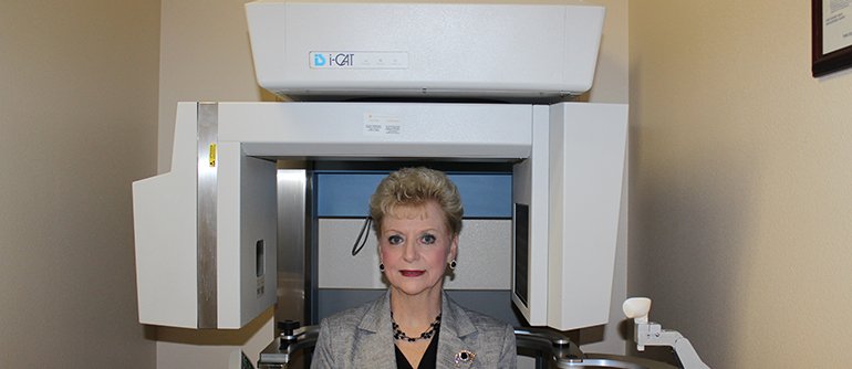
Computed tomography (CT) imaging, also referred to as a computed axial tomography (CAT) scan, involves the use of rotating x-ray equipment, combined with a digital computer, to obtain images of the body. Using CT imaging, cross sectional images of body organs and tissues can be produced. Though there are many other imaging techniques, CT imaging has the unique ability to offer clear images of different types of tissue. CT imaging can provide views of soft tissue, bone, muscle, and blood vessels, without sacrificing clarity. Other imaging techniques are much more limited in the types of images they can provide.

Cone Beam CT is used by Dr. Letelier to improve diagnosis and treatment planning in the following cases:

  • Dental implants
    • Location of anatomic structures: mandibular canal, submandibular fossa, incisive canal, maxillary sinus
    • Size and shape of ridge, quantity and quality of bone
    • Number, orientation of implants
    • Need for bone graft, sinus lift
    • Use of implant planning software
  • Oral and maxillofacial surgery
    • Relationship of third molar roots to mandibular canal
    • Localization of impacted teeth, foreign objects
    • Evaluation of facial fractures and asymmetry
  • Oral and maxillofacial pathology
    • Localization and characterization of lesions in the jaws
    • Effect of lesion on jaw in 3rd dimension: expansion, cortical erosion, bilateral symmetry
    • Relationship of lesion to teeth and other structures